Cow anatomy
The cow is one of the oldest domesticated animals. For many centuries, the most diverse breeds of cows have been bred, but all have the same anatomy. This article details the structure of the body and internal organs of these animals.
Head
It has a massive appearance, slightly elongated shape with an oblong face and a wide forehead. The skin is covered with short hair. On the head are eyes, nose, mouth and ears.

Skull
The cranial bones of cows are very durable.
Their skull can be divided into two departments:
- cerebral - a cavity for the brain, formed from 7 separate bones;
- front - the front of the head (muzzle), on which the eyes, nose and mouth are located.
Learn more about the structure of the skull of a cow. The calf has almost the same size of both of these parts. But in the process of growing up, the front part gradually becomes larger than the brain and stretches forward.

Eyes
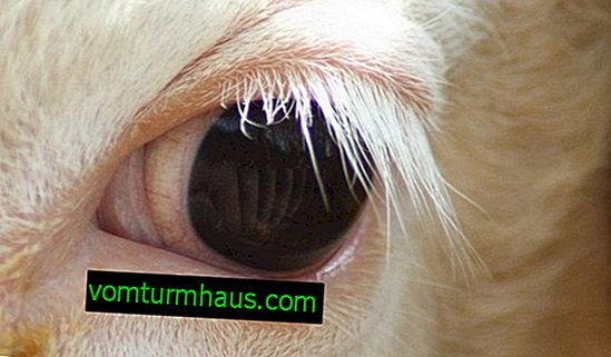
Cows have large eyes located on the front of the skull symmetrically to each other. Lateral vision in animals is monocular, and central - binocular. This means that they perceive the image located on the side of the head with only one eye, and the image located in front of them with two eyes. Using binocular vision, the cow sees both the ground directly in front of him and the terrain, but does not distinguish between the details. Important! Cows see the world around them on an enlarged scale and poorly distinguish colors. Eyeballs are located in special cavities on the surface of the head, orbits, and consist of such elements:
- Outer sheath - includes the cornea and sclera. Muscle tendons are attached to the latter, holding the eye inside the orbit. The cornea consists of many small nerves and provides light to the inner lining of the eye.
- Middle shell - consists of an iris with a small pupil, a thin shell of blood vessels and regulating the degree of convexity of the ciliary lens.
- The lens - has the form of a focusing image curved from two sides of the lens.
- Vitreous - supports the round shape of the eye, promotes metabolic processes and conducts rays of light.
- The inner shell (retina) - it displays the image seen by the animal, and then is converted into a nerve impulse transmitted to the brain.

Teeth
In cattle, they are arranged in such a way that they can only chew on plant foods. In the mouth of newborn calves, 20 milk teeth are placed; in the process of growing a calf, they gradually fall out and are replaced by permanent teeth. This process ends when the animals are 1.5 years old. Adult cows, like humans, usually have 32 teeth.
We advise you to learn about the number, structure and causes of tooth loss in a cow. On the lower jaw there are 8 incisors that capture and cut grass. They are quite sharp and are located at a slight slope forward.
Cutters have different names:
- hooks - located in the center;
- internal averages - located on both sides of the hooks;
- external averages - located on both sides of the internal averages;
- patches - located behind the outer middle incisors on two opposite sides of the jaw.
The cow captures the greens with the help of lips and a rough tongue and tightly presses the bunch to the incisors. Then she sharply moves her head and the grass is carefully cut, leaving roots in the ground.

The ears
The auricles of animals are large, composed of elastic cartilage and muscles, so cows often move their ears in different directions. Their shape looks like a horn. The outer part of the auricle is covered with short hair, and the inside is long. The inner ear consists of a tympanic membrane, a malleus, and an anvil.
Did you know? Cows can distinguish between the sounds of musical instruments and react differently to each of them.
Animals have very good hearing and ability to remember voices. They recognize sounds with frequencies up to 5000 Hz and can memorize them.

Horns
Two horns are located on the frontal bones of a cow’s head. Their surface is solid and formed by the epidermis of the horn, and the inside is hollow. Cow horns can stick up or to the sides. Their growth directly depends on the metabolic rate in the animal body. Find out also why a cow needs horns. Horns are not a mandatory attribute of cows. As a result of selection, hornless livestock breeds, which are called hornless, were also bred.

Skeleton
It is characterized by massiveness and durability. Bones easily support the weight of muscles and large internal organs. You will be interested to read more about the skeleton of a cow. The skeleton of a cow consists of two parts:
- axial - the skull, chest and spine belong to it;
- peripheral - includes the limbs of the animal.
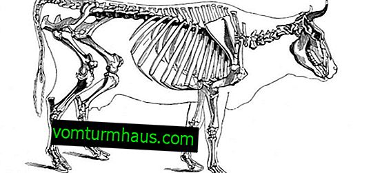

Spine
It stretches along the entire body of the cow, starting from the head and ending with the tail.
The spine consists of several sections:
- The cervical region is the area located between the skull and the beginning of the chest. It consists of 7 durable vertebrae that provide head movement. They help the cows for a long time to keep their head bowed to the ground and not feel tired.
- The thoracic part is the longest section of the spine. Located behind the cervical spine and stretches to the lower back. It consists of 13 vertebrae to which the ribs of the animal are attached. Together, these bones form the rib cage. Ribs located near the lower back cover the lungs of the animal and move noticeably during breathing.
- Lumbar - consists of 6 vertebrae.
- Sacrum - includes 5 vertebrae.
- The tail section - has from 18 to 20 mobile vertebrae.

Limbs
Cows have two pairs of legs growing from the lower torso. The front and hind limbs have a different structure.
Important! From the point of view of anatomy, not only the legs themselves, but also the bones connected to them belong to the limbs of cows.
According to their location, the limbs are divided into two types:
- chest - are in front; one limb contains a scapula, shoulder, forearm, and hand. The brush is divided into the wrist, metacarpus and fingers;
- pelvic - located behind; one limb consists of the pelvic bone, tubular femur, lower leg and foot.

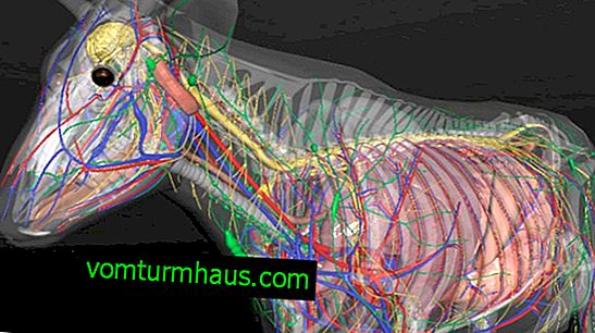
Internal organs and systems
The general vital activity of the animal is carried out thanks to the coordinated work of all systems of internal organs. These systems combine individual organs to perform a specific function.
Muscular
Cows have well-developed muscles and look strong. In the total weight of an adult animal, muscle weight is almost half. The entire musculature of the body is divided into muscles of the head and muscles of the body.
The muscles of the head include:
- facial muscles - are responsible for the “facial expressions” of the muzzle, give animals the opportunity to open their mouth and eyes, move their lips and move their nostrils;
- chewing muscles - fully correspond to its name and provide the possibility of movement of the jaws.
Did you know? Cows feel the Earth’s magnetic field well and prefer to stand so that their torso is parallel to its lines of force.
Torso muscles are also divided into several groups:
- shoulder muscles - provide a connection between the front legs of the cow and the humerus, are responsible for the movement of the forelimbs;
- sternum muscles - have a solid base and are located on the edges of the animal. They hold the internal organs inside the body. During inhalation, they stretch and enlarge the chest so that the cow can fill the lungs with air, and during the exhalation, it is narrowed;
- vertebral muscles - include many individual muscles responsible for the movement of the head, neck and tail. They also provide mobility of the lower back and spine, lifting the pelvis;
- muscles of the abdominal cavity - are able to contract and relax; during compression, they exert pressure on the internal organs of the cow, contributing to various vital processes (urination and defecation, childbirth, vomiting, or burping).

Nervous
Each part of the nervous system consists of various elements responsible for the performance of a certain function.
- The brain . It is the main organ of the central nervous system. It is located inside the skull and is divided into two hemispheres, covered with bark with convolutions. Between these two parts passes a dividing groove. The brain controls the entire life of the cow; its mass can reach 550 g.

- Spinal cord Belongs to the central nervous system, located inside the spine in the hollow channel. The spinal cord is divided into several sections and is responsible for performing unconditioned reflexes (limb movement, breathing, etc.). Its length can reach 180 cm.
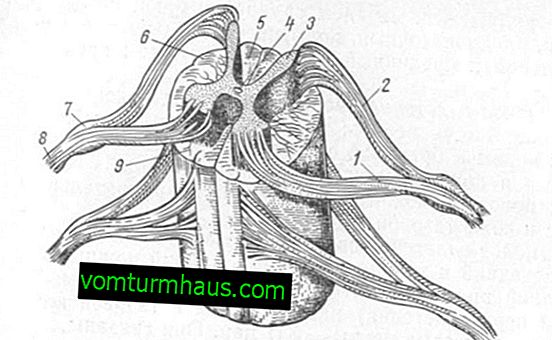
- Peripheral nervous system . It consists of many nerves connecting the brain and spinal cord with muscles, blood vessels, membranes of the abdominal cavity and excretory glands.
- Autonomic nervous system . It is a set of centers located in the brain (control the work of the pupil, lacrimal and salivary glands, lungs and pelvic organs) and in the spinal cord (provide the work of internal organs of the abdominal cavity and smooth muscles of blood vessels), and in addition, small nerve nodes scattered throughout the body.


Breathing
In cows, it is represented by a pair of well-developed lungs located in the chest under the ribs. The device of the animal’s lungs is similar to that of a human. At the time of inspiration, the light cows expand under the influence of internal pressure and are filled with air. The exhalation is carried out using the muscles of the sternum and diaphragm, which compress the lungs and squeeze air out of them.

Cardiovascular
This system in cows consists of a heart and a set of vessels - veins and arteries.
The heart is a muscle divided into two parts and consisting of four chambers - two atria and two ventricles. The heart muscle of cows is large and strong; in a day it pumps several tons of blood through itself. Blood is transported to all organs and tissues of the body, saturating their cells with necessary oxygen, various nutrients and water.
In the arteries, blood moves from the heart to the rest of the body, and through the veins, giving oxygen and nutrients, comes back.

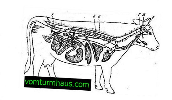
Digestive
It has a large length and multi-component structure. It provides the digestion of hard food, supplying the body with the substances necessary for a full life. Read also about the structural features of the digestive system of a cow.
- The oral cavity . Carries out capture of food with the help of lips and tongue and its further chewing with plentiful salivation.
- Esophagus . Connects the oral cavity to the stomach.
- The stomach . Consists of a grid, a scar, a book and an abomasum. The grid filters food by passing only well-ground semi-liquid gruel from the feed through it. She sends poorly chopped pieces of food for digestion into the rumen or into the esophagus to return to the mouth and chew more thoroughly. Gastric juice is produced only in the abomasum, it is here that the final breakdown of food occurs.
- Pancreas It secretes gastric juice and bile, which digest food.
- The small intestine . Connected with abomasum, food enters it to absorb the necessary nutrients.
- Large intestine . The last element of the digestive system of cows consists of three main departments. In the cecum, food is fermented. After this, the mass enters the colon, where excrement is formed. The latter are removed from the body of the animal to the outside through the small anal opening located under the tail.

Urinary
In the body of cows, the process of formation, accumulation and excretion of urine is carried out using the following organs:
- kidneys - urine is formed in them;
- ureters - transport urine from the kidneys to the bladder;
- bladder - collects urine, after filling it moves to a special channel;
- urethra - connected to the bladder, through it the urine leaves the body.
Did you know? An adult cow excretes from 6 to 20 liters of urine within 24 hours.
Genitals
The reproductive system is responsible for the reproduction of animals. It is represented by reproductive organs, which in cows and bulls differ in structure.
Cows
The reproductive organs of a cow are involved in fertilization, gestation and childbirth.
The reproductive system of the animal includes such elements:
- vulva - a hole under the anal;
- clitoris - increases uterine contractions, helping to cause additional stimulation in the cow;
- shameless lips - folds located at the entrance to the vagina;
- vagina - connects the external opening of the vulva with the uterus, serves to perform copulation;
- uterus - consists of muscle tissue, a fertilized egg is placed in it, which eventually turns into a fetus;
- fallopian tubes - here the egg is fertilized;
- ovaries - they contain eggs.

Bull
The genitals of the bull produce sperm, copulate with the cow and fertilize her eggs.
These include:
- penis - intended for urination and the introduction of seminal fluid into the vagina of the female;
- prepuce - in a relaxed state, covers the outer edge of the penis, and during an erection opens it;
- urogenital canal - connects the bladder to the top of the penis, removes sperm and urine;
- seed tube - provides transportation of seminal fluid to the place of fertilization;
- spermatic cord - a fold in the abdominal cavity containing nerve endings, blood vessels, and the vas deferens;
- testes (testes) - are responsible for the production and storage of sperm;
- scrotum - a large fold of skin inside which the testes are placed.

The structure of the udder
The udder has a complex structure and is located in the lower abdomen of a cow. It consists of four lobes, each of which has a nipple. The skin of the udder is elastic and dotted with short hairs.
The udder of an animal consists of several types of tissue:
- glandular - is a cluster of alveoli, inside of which milk is produced. Through a network of ducts and canals, milk from the alveoli enters the nipples;
- connective - located around the alveoli and is penetrated by blood vessels, nerve endings and lymph nodes, performs a protective function;
- fatty - protects the udder from external irritants.
The nipple on the udder is the muscle fold through which the nipple canal passes. On it the output of milk is carried out.

Tail
The long spine of a cow ends with a tail. It consists of movably connected vertebrae, and at the end of the tail there is a small brush. Cows use their tail to scare away annoying flies, horseflies and mosquitoes.

The internal structure of the cow as a whole is the same as that of other warm-blooded animals, but it also has some features. Знание анатомии этих животных помогает в уходе за ними и в лечении заболеваний.